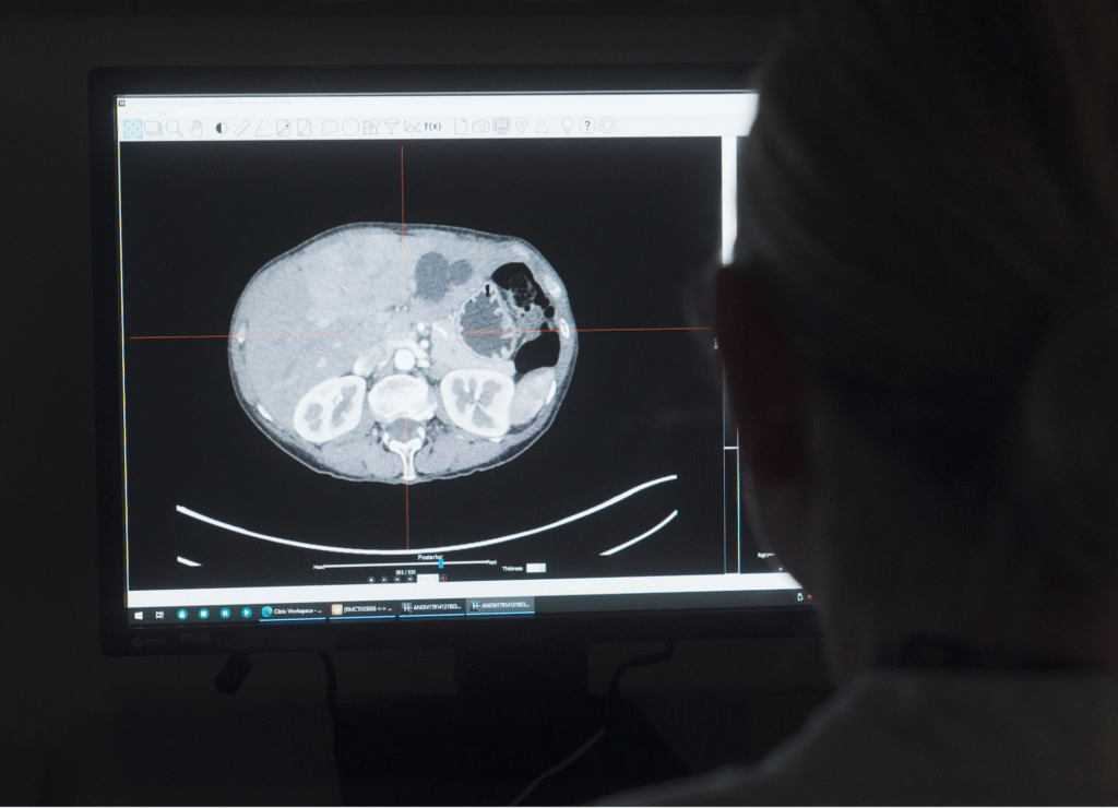
NoloSight’s ProPET is an advanced probabilistic reconstruction and image analysis methodology specifically designed to enhance lesion detection in PET/CT imaging. By combining informed prior models with machine learning, ProPET improves the ability to identify small lesions while maintaining clinical accuracy.
How It Works: The ProPET Framework
ProPET applies a probabilistic approach to PET image analysis, that incorporates three key components:
Prior Information
Expert knowledge about tracer distribution patterns in specific clinical contexts. Rather than relying on generic filtering, ProPET uses informed prior models that reflect the biological reality of disease—such as an expected sharp contrast between high-uptake lesions and surrounding normal tissue in cancer imaging.
Scanner Physics
Accurate models of the point spread function and spatially correlated noise characteristics. ProPET is calibrated for each specific PET scanner platform. This scanner-specific approach ensures the methodology works optimally across different imaging systems.
Machine Learning
Neural networks trained to efficiently estimate posterior statistics across whole-body scans. Rather than computationally expensive sampling methods, ProPET uses machine learning to rapidly compute posterior mean activity, uncertainty estimates, and lesion probability maps in real-time.
Result: This probabilistic framework enables real-time clinical application while maintaining theoretical rigor, providing richer diagnostic information than conventional SUV-based analysis alone.


Enhanced Lesion Detection
ProPET identifies additional suspicious foci across all anatomical regions, particularly excelling at detecting small lesions that may be obscured by noise and smoothing artifacts in conventional reconstruction. In clinical evaluation, ProPET consistently identified more lesions while missing none that were detected with standard reconstruction.
Improved Diagnostic Confidence
The posterior probability distributions generated by ProPET provide uncertainty estimates and lesion probability maps at each voxel, enabling clinicians to distinguish confident findings from borderline cases. This enhanced confidence supports more decisive clinical decision-making.
Seamless Clinical Integration
ProPET is designed to work with standard PET/CT acquisition protocols and scanner hardware. It processes data retrospectively from existing imaging systems without requiring hardware modifications or changes to scanning procedures.
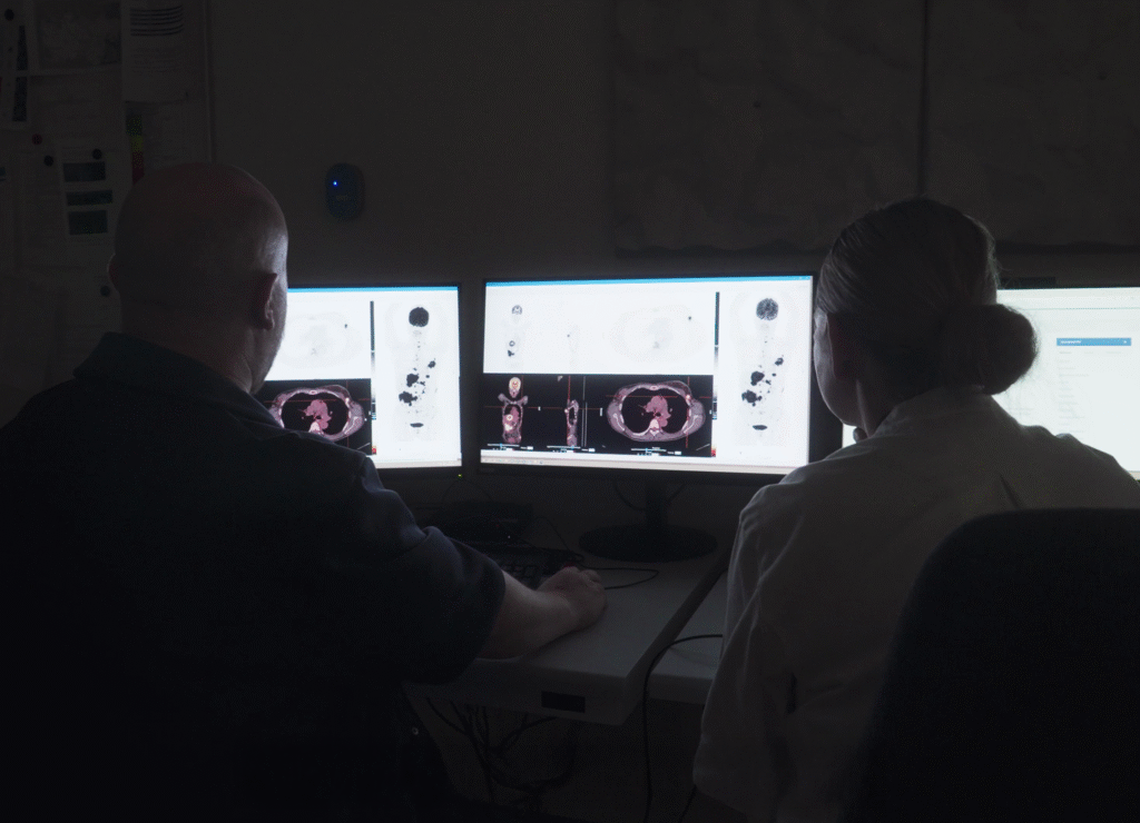
Close collaboration with medical professionals—particularly nuclear medicine specialists and oncologists—has been instrumental in shaping ProPET’s development. Working directly with clinicians has provided invaluable insight into:
This partnership has ensured that ProPET addresses genuine clinical problems and integrates naturally into existing practice. Feedback from practitioners continues to guide refinement of the technology, ensuring it delivers meaningful improvements where they matter most—at the point of care.
ProPET’s development has focused on FDG-PET/CT imaging in oncology, with particular emphasis on improving detection of:
Future work aims to expand clinical evaluation to additional oncological applications and explore potential use in other disease areas where improved lesion detection would have clinical value.
✓ Enhanced detection of small lesions in PET imaging
✓ Improved diagnostic confidence through probability estimates
✓Better characterization of lymph nodes and metastatic disease
✓ Integration with standard clinical workflows
✓ Multiple scanner platform compatibility (with platform-specific calibration)
Prospective clinical studies are underway to:
The NoloSight team remains committed to rigorous validation and evidence-based development, ensuring ProPET delivers reliable clinical value as it advances toward broader implementation.
While originally designed to enhance PET scan imaging, the technology is also compatible with other modalities such as MR and MRI. Its adaptable algorithm improves image clarity across various scan types, enabling earlier and more accurate identification of a wide range of conditions.
This includes not only cancer but also complex neurological disorders like Parkinson’s disease and Alzheimer’s. The versatility of the system opens new possibilities for diagnosis, monitoring, and research across multiple fields of medicine.